Earlier this year, ACA member Kenneth Weber, DC, PhD, was awarded two grants totaling more than $5 million from the National Institutes of Health (NIH). Dr. Weber, a chiropractor and neuroscientist, is a senior research scientist in the Division of Pain Medicine at Stanford University School of Medicine. With his current research, he aims to use MRI (magnetic resonance imaging) to find better diagnostic and predictive information for spinal cord conditions. Grant funding will also allow Dr. Weber to build a multidisciplinary research team, which he hopes can include chiropractic researchers.

“It is an important goal of mine to help train and mentor chiropractic researchers, so they can have successful research careers and grow chiropractic research,” he explained. “This funding can be an opportunity for more chiropractors to get research training.”
The grants, funded by the National Institute of Neurological Disorders and Stroke (NINDS) of the NIH, will help pave the way for chiropractic opportunities in research, as similar funding already has for Dr. Weber. “It takes time to get to the point where you can successfully compete for these awards, and I am where I am today because of my previous mentors,” he said. “These funds are now allowing me to build my own team, and I am here to return the favor and help more chiropractors become independent researchers.”
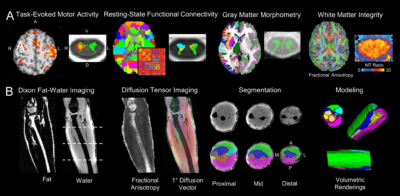
Over the next five years, Dr. Weber and his team will use MRI to explore motor and sensory processing. “The overall scope of these projects is to identify better markers of sensory and motor function, including pain, in spinal conditions with neurological injury,” he said. “There’s a large imaging component to our research, and one of the main techniques is MRI, which we use to assess brain, spinal cord, and muscle health.”
The first project, titled “MRI-Derived Neuromuscular Signatures to Predict Surgical Response in Degenerative Cervical Myelopathy,” is focused on degenerative cervical myelopathy (DCM), the most common form of spinal cord injury in adults, often older adults. This condition occurs when degenerative changes in the spine compress the spinal cord and cause injury to it, which can lead to pain and decreased muscle function in the limbs. Surgery is often a necessary treatment for this condition, Dr. Weber explained, but it can be difficult to know how older adults may respond.
“We don’t have a lot of good markers that predict surgical response,” he said. “The other concern is that these are older adults, that may have additional comorbidities, and we want to reserve surgery for those people who will respond.”
This study will use MRI of the brain, spinal cord and forearm muscles to develop markers of hand function in patients with DCM. Although the primary injury occurs in the spinal cord, Dr. Weber explained that there is evidence that the injury can extend to the brain and muscle. “By looking at the brain, spinal cord and muscle in a single study, we can get a more comprehensive assessment of the neuromuscular system,” he said. “[We can] use this information to potentially better grade the severity of DCM and predict surgical success.”
The hypothesis is that the patients with the injury limited to the spinal cord, and fewer changes in the brain and muscles, will respond better to surgery — “Their brain and muscles will be able to better recover hand function after the compression of the cord is removed,” Dr. Weber explained.
The second project is titled “Dermatomal Mapping with Spinal Cord Functional Magnetic Resonance Imaging.” In this research, Dr. Weber aims to better understand sensory processing in the spinal cord and test hypotheses related to dermatomal maps, which are common tools used in clinical practice. The research will involve stimulating different parts of the hands and looking at where the corresponding activation occurs in the spinal cord.
“In the brain, we know that there are differences in sensory activity depending on the side of stimulation,” he said. “Sensory activity in the brain is slightly different when you feel with your dominant hand versus your non-dominant hand — do these sidedness differences already appear in the spinal cord? The other aspect is sex differences. We know that there are large sex differences in the brain — do we see these sex differences showing up in the spinal cord, which is much earlier in the sensory processing, before the signals arrive in the brain?” Similarly, the project will look at how age affects sensory processing in the spinal cord.
The dermatomal mapping project will also explore how nerve injury affects sensory processing in the cord and the sensory activation maps. “We’ll be imaging patients with cervical radiculopathy, who have nerve root injury, and assessing how the sensory activity changes,” Dr. Weber said. “If you have injury to a nerve root, it’s expected that you’re going to have decreased sensation at that nerve root level, but how sensory activation changes in the other nerve roots that are still intact is largely unknown in the spinal cord.” Ultimately, Dr. Weber hopes that this information will improve the treatment of cervical radiculopathy.
A shared goal for both projects is to make MRI more quantitative. “Generally, when you have an MRI done for clinical purposes, you’ll have a radiologist read the images and write a narrative report based on what they see in the images,” he explained. “It tends to be more qualitative in that they’re viewing the images and giving you a verbal report. We are focused on making MRI more quantitative. There’s a lot more information in the images than what you can visually assess, and we can use different techniques to extract that information, which we hope will eventually allow us to be more quantitative in the diagnosis and prognosis of spinal conditions.”
Dr. Weber is hopeful about the impact that this research could have on the treatment of spinal conditions. While these grants are more focused on surgery, the next steps of this research will be to expand to non-surgical treatments, including chiropractic care. He’s excited to build a multidisciplinary team to move these projects forward. So far, there are physicists, pain medicine specialists, statisticians, neuroscientists, neurosurgeons and physical therapists collaborating in the research. “Hopefully, we can bring more chiropractors onto the research team in the near future,” he said.
If you have questions about Dr. Weber’s research, including an interest in research training, feel free to reach out to him at [email protected].